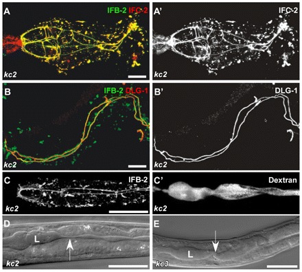
Fig. 5.

In ifo-1 mutant worms, IF proteins IFB-2 and IFC-2 colocalize and co-distribute with the CeAJ marker DLG-1. The intestinal lumen is perturbed but still tight. (A,A′) Fluorescence of dissected adult ifo-1(kc2) intestines detecting IFB-2::CFP and anti-IFC-2. IFC-2 colocalizes with IFB-2 in cytoplasmic aggregates and at the CeAJ in anterior intestinal cells. Note the presence of IFC-2 but not of IFB-2 in the adjacent pharynx. (B,B′) The re-distributed anti-IFB-2 immunofluorescence colocalizes with anti-DLG-1 immunofluorescence at the CeAJ but shows a more extensive staining adjacent to the junction and in cytoplasmic granules. (C,C′) Fluorescence micrographs depict the disturbed IFB-2::CFP distribution in kc2 mutant worms (strain BJ133) together with TRITC-labeled dextrane that was taken up by feeding. Note that the dextrane is restricted to the altered lumen indicating that the junctional seal is still intact. (D,E) DIC micrographs reveal an abnormal lumen (L) with multiple constrictions (arrow) and dilations in kc2 and kc3 mutant worms (strains BJ133 and BJ134, respectively). Scale bars: 10 μm in A,B; 100 μm in C-E.
