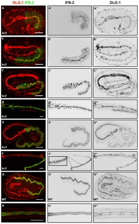
Fig. 6.

IFB-2 mislocalization in kc2 and kc3 mutants develops differently. Embryos (A-C,E,G) and L1/L2 larvae (D,F,H) were stained with antibodies against IFB-2 (green, A-H; inverted, middle column) and DLG-1 (red, A-H; inverted, right column). (A-C″) In the 1.5-fold stage of kc2 mutants, IFB-2 is localized at the apical part of intestinal cells in a punctate pattern (A-A″), starts to accumulate at the CeAJ at the 2-fold stage but is still present in apical puncta (B-B″) and becomes fully recruited to the CeAJ at the 3-fold stage (C-C″; note increase of cytoplasmic IFB-2 granules in number, especially in the anterior intestine). (D-D″) In kc2 L2, the aggregates further increase and are more evenly distributed throughout the intestine. (E-E″) By contrast, IFB-2 is distributed normally in kc3 embryos (compare with Fig. 3D). (F-F″) In the kc3 L1 stage, small gaps (see magnification in F’) appear in the apical IFB-2-positive network. The gaps increase in size in older worms until the phenotype resembles that of kc2 worms (see Fig. 1). (G-H″) In WT embryos and L1 larvae, the contiguous anti-IFB-2 and anti-DLG-1 signals delineate the endotube and the CeAJ, respectively. Scale bars: 10 μm.
