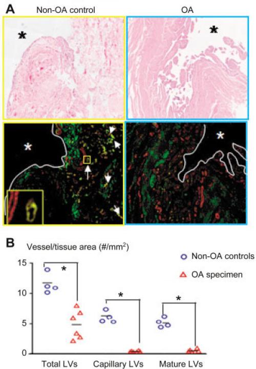
Figure 6.
Decreased lymphatic vessel formation in joints from patients with OA. Knee joint samples from patients with severe OA who underwent total knee arthroplasty (n = 6) or from patients without OA who had a fresh subcapital fracture of the femoral neck and had undergone total hip arthroplasty or hemiarthroplasty (n = 4) were used. A, Sections were stained with hematoxylin and eosin and double stained with anti-podoplanin antibodies (green) and α-smooth muscle actin (α-SMA) antibodies (red) and scanned as described in Figure 1. Photomicrographs show numerous lymphatic capillaries (green) and mature vessels (yellow) (white arrows) in the subsynovial area of a representative non-OA sample. A podoplainin+/α-SMA+ mature lymphatic vessel adjacent to a blood vessel (podoplanin–/α-SMA+) (boxed area) is shown at higher magnification in the inset. Asterisks indicate synovial space. Original magnification × 40. B, Histomorpho-metric analyses of lymphatic vessel density were performed. Each symbol represents a single sample; bars show the mean. * = P < 0.05. See Figure 3 for other definitions.

