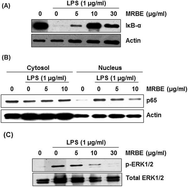
Figure 2.

Effect of MRBE on IκB-α degradation (A), p65 nuclear translocation (B) and ERK1/2 phosphorylation (C) in LPS-stimulated RAW264.7 cells. RAW264.7 cells were pre-treated with MRBE at the indicated concentrations for 2 h and then co-treated with (1 μg/ml) for 15 min (for Western blot of IκB-α and ERK1/2 phosphorylation) or 30 min (for Western blot of p65). DMSO was used as a vehicle. Cell lysate were resolved by SDS-PAGE, transferred to PVDF membrane, and probed with antibodies against IκB-α, p-ERK1/2, total ERK1/2 and p65. The proteins were then visualized using ECL detection. Actin was used as an internal control.
