Fig. 1.
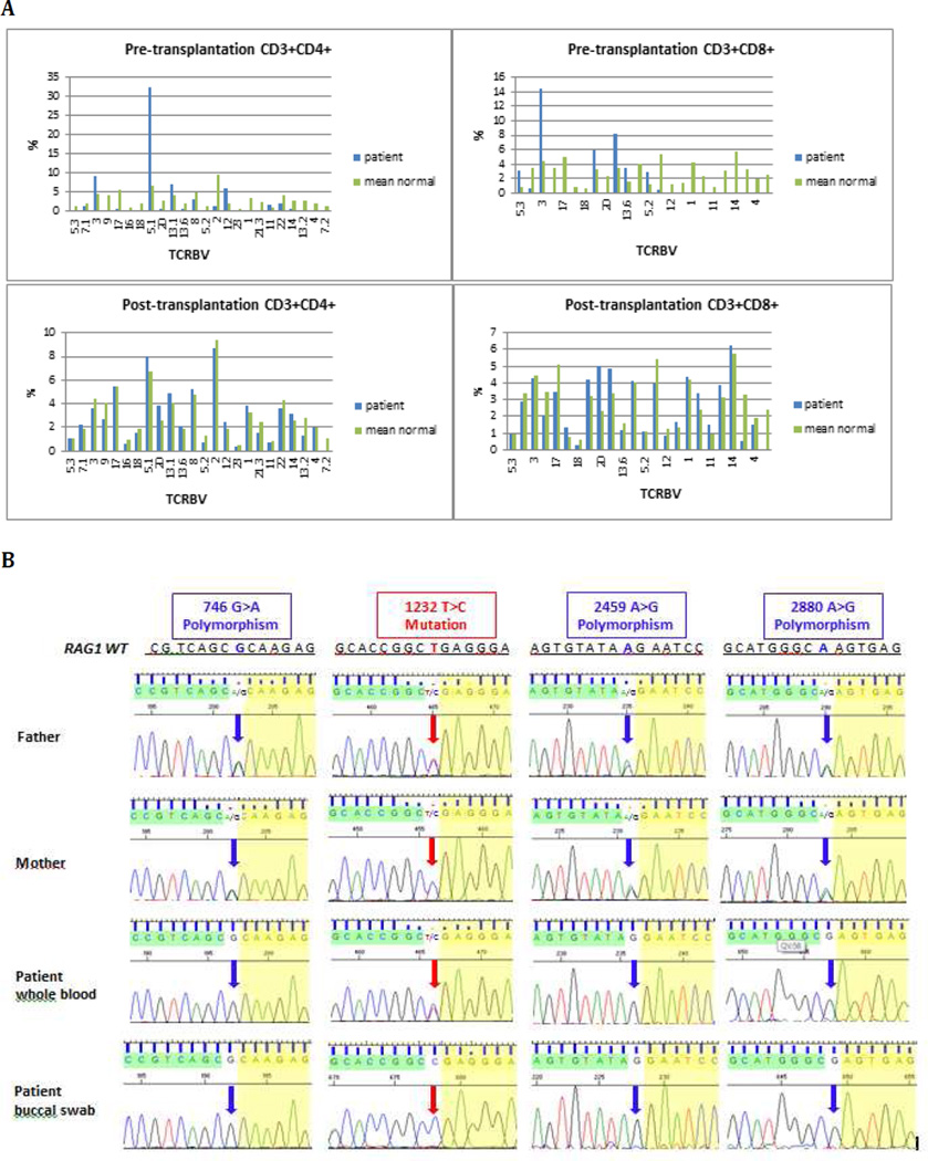
A TCRBV repertoire pre- and post-transplantation. Flow cytometric analysis of the percentage of CD4+ (left panels) and CD8+ (right panels) lymphocytes expressing the various TCRBV families before (top panels) and after (bottom panels) hematopoietic cell transplantation in the patient (blue bars), as compared to mean values from healthy age-matched controls (green bar)
B Results of RAG1 sequencing in the patient and his parents Chromatograms of Sanger sequencing demonstrating apparent heterozygosity for the 1232T>C mutation in patient’s whole blood and in both parents. Homozygosity for the same mutation is present in patient’s buccal swab DNA. Analysis of three known RAG1 polymorphisms demonstrates homozygosity in the patient’s samples, and heterozygosity in both parents.

