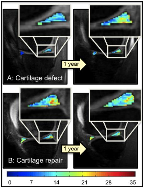
Figure 5.

T1ρ color maps of the anterior and posterior horn of the medial meniscus of 1 year and 2 year follow-up time-points, overlaid with the first-echo images. Superior: Control subject with cartilage defect at the medial femoral condyle, who did not receive a cartilage repair (CR) procedure. Inferior: CR subject with osteochondral transplantation at the medial femoral condyle. Blue color indicates low, red color high meniscus T1ρ values. Subjects with untreated cartilage lesion showed a higher increase in T1ρ values over time compared to the subject with CR.
