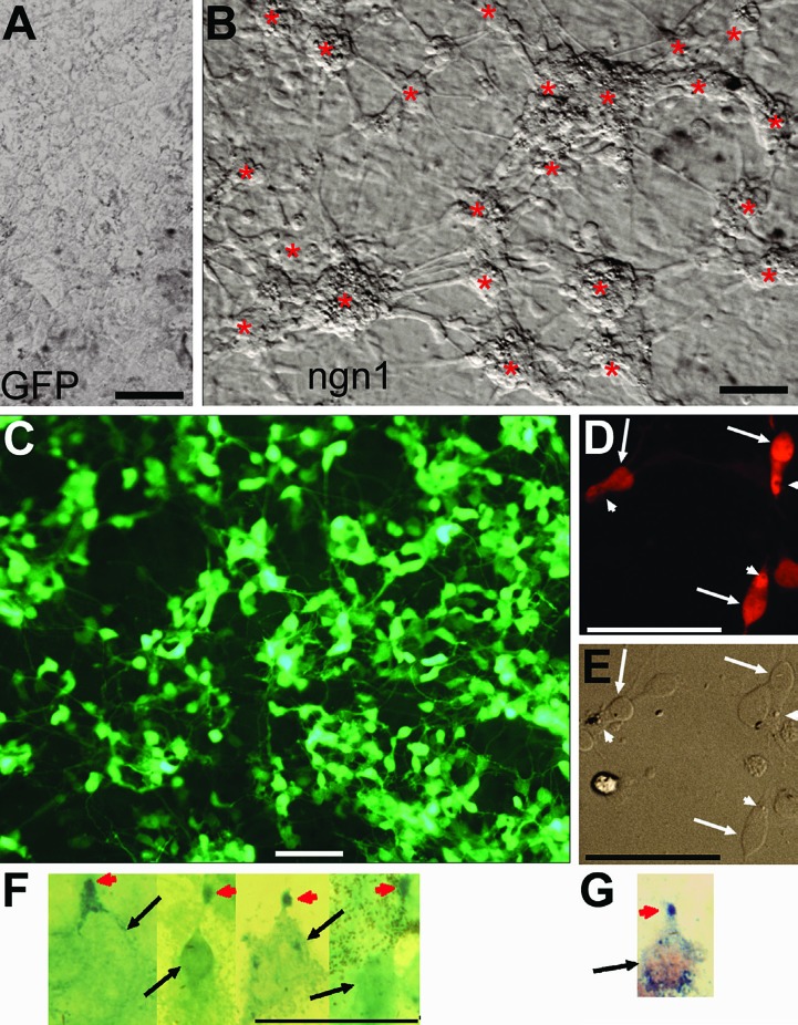
Figure 1.
Photoreceptor-like cells in reprogrammed RPE cell culture derived from day 6 chick embryos. A: Bright field view of a control culture infected with RCAS-GFP. B: Bright field view of ngn1 reprogrammed culture (infected with RCAS-ngn1) displaying neuron-like clusters (*), which were absent in the control (A). C: Epi-fluorescence view of reprogrammed culture immunostained for photoreceptor protein visinin. D, E: Morphologies of visinin+ cells viewed with bright field (E) and epi-fluorescence (D). Arrows point to the cell body and arrowheads point to a structural feature reminiscent of the lipid-droplet typically present in chick photoreceptors. F: Morphologies of red opsin+ cells in ngn1 reprogrammed culture. Arrows point to cell bodies. Arrowheads point to cells’ apices decorated by anti-red opsin immunostaining. G: A cell double-labeled for visinin (in red) and red opsin (in blue) in ngn1 reprogrammed culture. Scale bars: 50 μm. [Data are from the authors’ laboratories]. [Modified from Yan et al., 2010. Originally published in Journal of Comparative Neurology; DOI 10.1002/cne.22236].67

