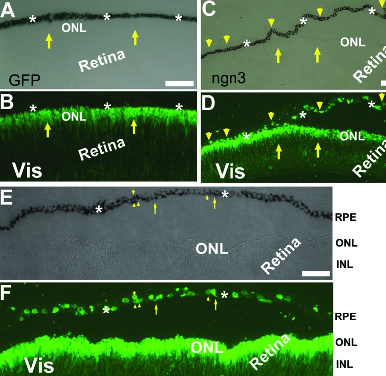
Figure 4.
Visinin+ cells in the RPE layer in chick embryos infected with RCAS expressing ngn3 (RCAS-ngn3). A, B: Bright-field (A) and epi-fluorescence of immunostaining for visinin (Vis, B) of an E7.5 control eye infected with RCAS-GFP. C, D: Bright-field (C) and epi-fluorescence of visinin immunostaining (D) of an E7.5 eye infected with RCAS-ngn3. E, F: Bright-field (E) and epi-fluorescence of visinin immunostaining (F) of a detached region in E7.5 eye infected with RCAS-ngn3. Arrows point to visinin+ cells of the retina, except in E,F, where arrows point to visinin+ cells with a neural-like process in the RPE layer. Arrowheads point to visinin+ cells in the RPE layers. ONL, outer nuclear layer; INL, inner nuclear layer [Data are from the authors’ laboratories]. [Modified from Li et al., 2010. Originally published in Investigative Ophthalmology & Visual Science; DOI:10.1167/iovs.09-3822].68

