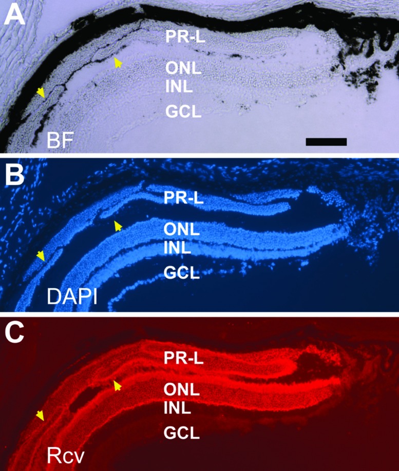
Figure 6.
Recoverin+ cells in the subretinal space in a 2.5-month-old PRPE65-ngn3 transgenic mouse. Shown are views of the same sample under bright field (BF, A), counterstaining with nuclear dye (DAPI, B), and immunostaining for photoreceptor protein recoverin (Rcv, C). Arrowheads point to darkly pigmented tissue associated with, as well as demarcating different domains of, recoverin+ (PR-L) cells. ONL, outer nuclear layer; INL, inner nuclear layer; GCL, ganglion cell layer Scale bar, 100 μm, applies to all panels. [Data are from the authors’ laboratories]. [Modified from Yan et al., 2013. Originally published in Investigative Ophthalmology & Visual Science; DOI:10.1167/iovs.13-11936].73

