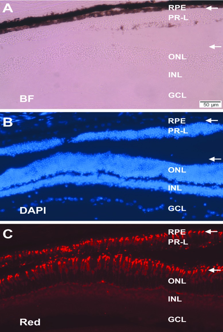
Figure 7.
Structural resemblance of PR-L cells to photoreceptors and the preservation of the RPE. Shown are histology and immunohistology of the retina from a 1-year-old PRPE65-ngn3 transgenic mouse under bright field view (A), DAPI counterstaining (B), or immunostaining for red opsin (C). In the aging animal, the single-layered RPE and a layer of PR-L cells were apparent. White arrows point to the immunostained outer segments of red cones in the ONL and the apices of similarly oriented PR-L cells. ONL, outer nuclear layer; INL, inner nuclear layer; GCL, ganglion cell layer. Scale bar, 50 μm, applies to all panels. [Data are from the authors’ laboratories]. [Modified from Yan et al., 2013. Originally published in Investigative Ophthalmology & Visual Science; DOI:10.1167/iovs.13-11936].73

