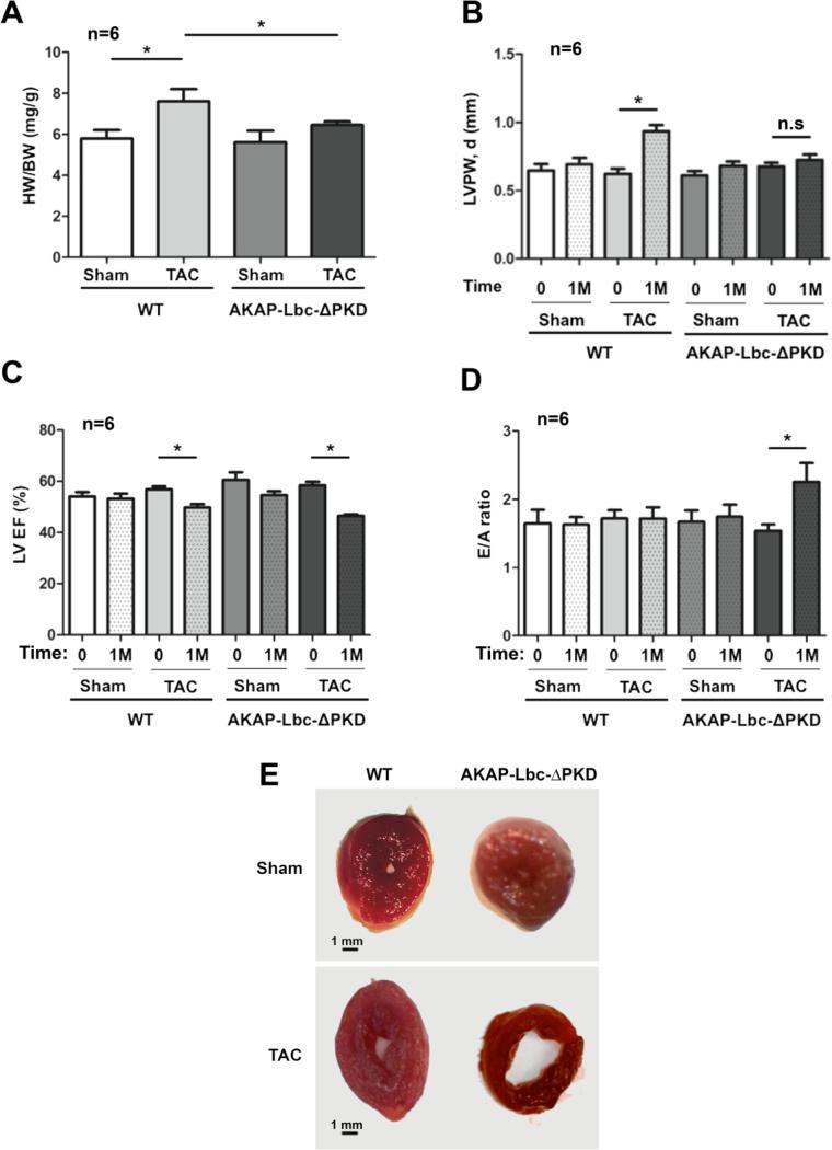
Figure 3. Echocardiographic and gravimetric characteristics of hearts from AKAP-Lbc-ΔPKD mice and control WT littermates, at baseline (0) and after TAC or sham surgery for 1 month (1M).
A) WT mice subjected to TAC for one month develop a significant increase in HW to BW ratio (*p < 0.01 WT/sham vs. WT + TAC); HW to BW ratio for AKAP-Lbc-ΔPKD mice + TAC is comparable to AKAP-Lbc-ΔPKD + sham. HW to BW ratio is increased in WT + TAC compared to AKAP-Lbc-ΔPKD + TAC (*p < 0.05). B) LV posterior wall thickness in diastole (LVPW,d) is increased in WT + TAC compared to baseline measurements (0), and AKAP-Lbc-ΔPKD mice + TAC. C) WT + TAC and AKAP-Lbc-ΔPKD + TAC show a decrease in LV Ejection Fraction (EF), compared to baseline (0) (*p < 0.05). D) E/A ratio, the early (E) to late (atrial; A) ventricular filling velocity ratio, is significantly increased in AKAP-Lbc-ΔPKD + TAC, compared to WT + TAC, sham and baseline (0) measurements (*p < 0.05). E) WT and AKAP-Lbc-ΔPKD heart section at the mid-papillary. In response to TAC, WT hearts develop concentric hypertrophy, whereas AKAP-Lbc-ΔPKD hearts exhibit a dilated LV chamber and thin walls.

