
Figure 2.
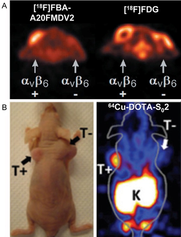
Small-animal PET imaging of integrin αvβ6 expression in cancer. A: Representative transaxial PET images of [18F]FBA-A20FMDV2 and [18F]FDG in the same mouse with the positive (αvβ6-expressing DX3puroβ6) tumors located near the left shoulder and the negative (control DX3puro) tumors near the right shoulder. Adapted with permission from the American Association for Cancer Research: ref. [50]. B: The photograph and PET image of a mouse bearing both BxPC-3 αvβ6-positive (T+) and αvβ6-negative 293 (T-) xenografts injected with 64Cu-DOTA-S02. Adapted with permission from the American Association for Cancer Research: ref. [59].