Figure 3.

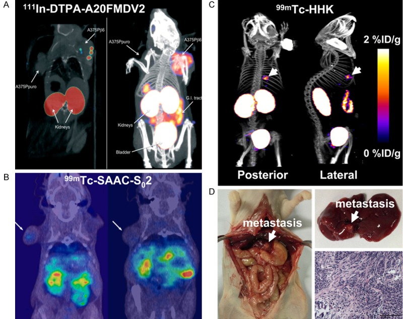
Small-animal SPECT imaging of integrin αvβ6 expression in cancer. (A) Small-animal SPECT (left) and SPECT/CT (right) images of mice bearing αvβ6-negative A375Ppuro (left shoulder) and αvβ6-positive A375Pβ6 (right shoulder) tumors with 111In-DTPA-A20FMDV2. Adapted with permission from ref. [71]. Copyright (2010) Pathological Society of Great Britain and Ireland. (B) Coronal SPECT/CT images of a mouse bearing αvβ6-positive HCC4006 xenograft (left) and a mouse bearing αvβ6-negative H838 xenograft (right) after injection of 99mTc-SAAC-S02. Adapted with permission from ref. [74]. Copyright (2014) American Chemical Society. (C) Whole-body posterior and right lateral SPECT/CT images of nude mice with liver metastasis of αvβ6-positive BxPC-3 tumors after 99mTc-HHK injection. (D) Mouse from (C) was sacrificed, and the liver metastasis was verified. This research was originally published in ref. [75]. Copyright (2014) the Society of Nuclear Medicine and Molecular Imaging, Inc.
