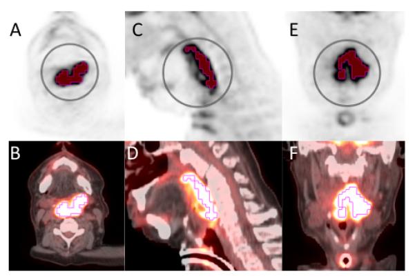
Figure 3.

58-year-old African American man with a greater than 60 pack year history of smoking, presented with progressive dysphagia and odynophagia. He was found to have a large left oropharyngeal mass. Biopsy was taken showing invasive moderately differentiated keratinizing squamous cell carcinoma that was negative for P16 and HPV. Staging PET scan with axial PET (A), axial fused (B), sagittal PET (C), sagittal fused (D), coronal PET (E), and coronal fused (E). The primary lesion based is shown with contouring based on threshold method at 50% of SUVmax. SUVmax 16.7, SUVmean 10.9, SUVpeak 13.7, MTVthreshold 35.0, TLGthreshold 382.7.
