
Figure 1. Structures of μ-conotoxins.
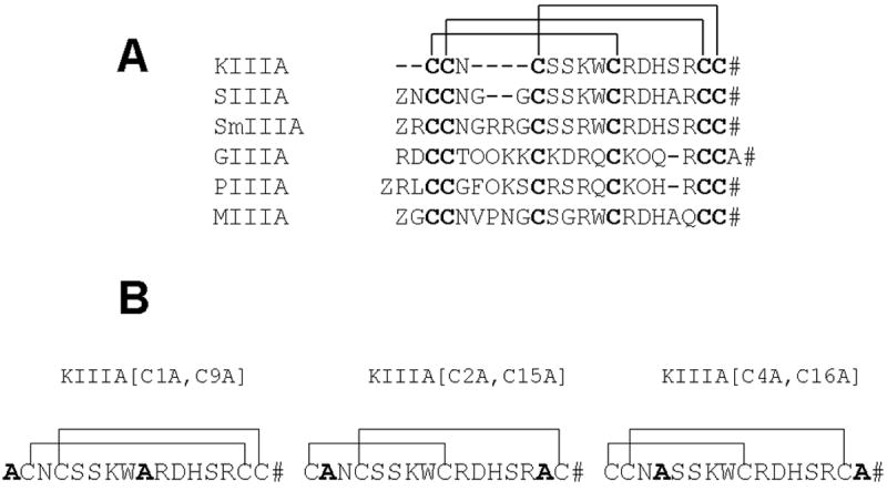
(A) – Alignment of sequences of several representative μ-conotoxins. The Roman numerals label the three intercysteine “loops.” (B) - Structures of the disulfide-deficient analogs of μ-conotoxin KIIIA studied in this report. #, amidated C-terminus; Z, pyroglutamate; O, hydroxyproline.