
Figure 7. Structural properties of the disulfide-deficient analogs studied by molecular dynamics.
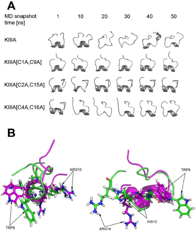
(A) - The total of 6 snapshot trajectories of 1, 10, 20, 30, 40 and 50 ns MD simulations of KIIIA and its analogs missing specific disulfide bridges, specifically KIIIA[C1A, C9A], [C2A, C15A] and [C4A, C16A]; (B) - Conformation of the KIIIA (green) and KIIIA[C1A, C9A] (magenta) backbone, shown as cartoon representation, carrying four pharmacophore residues (W8, R10, H12, R14) indicated by arrows. End-on view (left) and side view (right) of the helical secondary structure are presented.