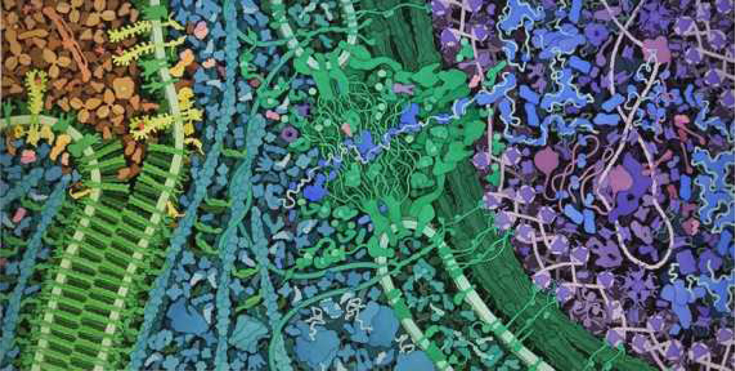
Figure 6.
The final molecular landscape depicting VEGF signaling. Blood serum is shown in tan at the upper left. The adherens junction between two cells is in green at left, with surface VEGF receptors shown in yellow. The cytoplasmic proteins are in turquoise, and the nuclear pore is at the center in green. The nucleus is at the right, with proteins shown in blues and purples.

