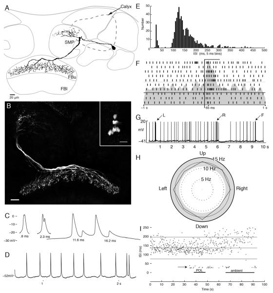
Figure 4.
Tangential neurons of the fan-shaped body. A: Camera lucida reconstruction of a tangential neuron with arbors in the superior protocerebrum (SMP) and branches that span a stratum of the upper division of the fan-shaped body (FBu). B: Confocal stack showing the morphology of eight tangential neurons filled by dye injection into a single neuron. Inset shows eight cell bodies brightly labeled. C: Examples of the double spike waveforms of various latencies. D: Example voltage trace showing regular spiking activity interrupted by doublets. E: Histogram of observed interspike intervals (ISIs). The doublets form a separate population of spikes with ISIs < 50 msec. F: Raster plot showing spiking activity during 10 repeats of a light flash presented from overhead. Bar indicates period of stimulus. G: Example voltage trace from one tangential neuron showing the change in firing activity in response to air puffs to the left (L) and right (R) sides and from the front (F) of the head. H: Direction preference plot for moving bar stimulus. This stimulus has no effect on this cells firing (Rayleigh test, n = 257, r = 0.006, P > 0.05). I: ISIs (ordinate) plotted as they occurred for 100 seconds of the experiment. Doublets (short ISIs) occurred at the moment of change of the stimulus modality. Scale bars = 20 μm.

