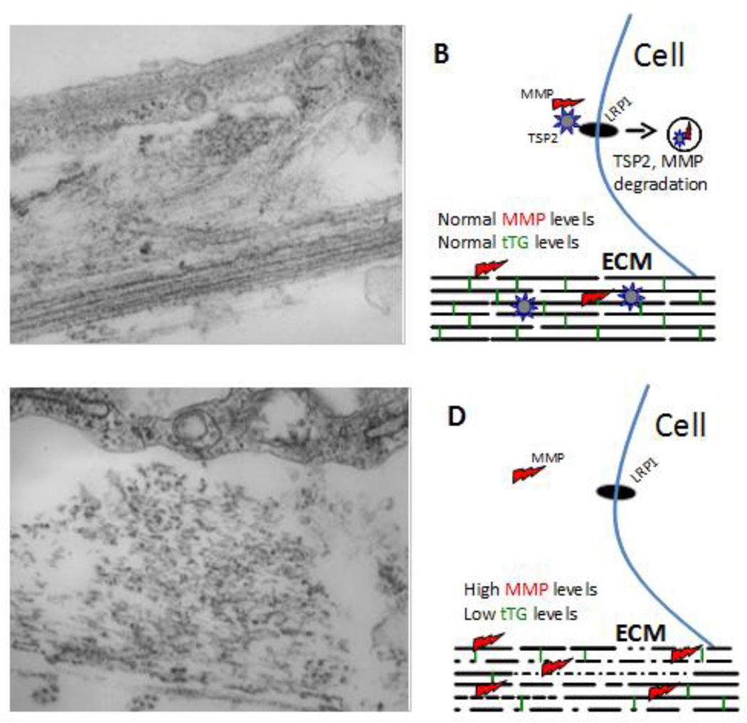
Figure 2. Role of TSP2 in ECM assembly.
Representative transmission electron microscopy images of WT (A) and TSP2-null (C) dermal fibroblasts cultured for 10 days in the presence of ascorbic acid are shown. WT cells display robust and organized deposition of collagenous ECM characterized by layers of highly dense and parallel collagen fibrils (arrow in A) as well as formation of dense fibril bundles (arrowhead in A). In contrast, TSP2-null cells display suboptimal deposition of discontinuous collagen fibrils (arrow in C) and disorganized bundles (arrowhead in C). Asterisks (*) denote cell areas. B, D show schematic representations of the interplay between TSP2, MMPs, and tTG in the process of ECM assembly. In WT cells, TSP2-MMP complexes interact with LRP1 and are targeted for intracellular degradation allowing for balanced MMP and tTG activity. In TSP2-null cells, due to the lack of TSP2 there is an increased accumulation of MMP2 resulting in degradation of collagen and the crosslinking enzyme tTG. As a result, ECM assembly is compromised due to reduced presence of crosslinks (green vertical lines in B and D) and changes in collagen deposition (gaps in collagen fibrils). Original magnification 4,000× (A, C).

