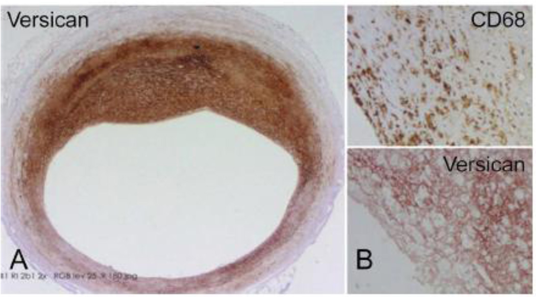
Figure 1.
A. A section through an early human coronary atherosclerotic lesion immunostained for versican illustrating marked accumulation of the ECM proteoglycan. B. Upper panel – These lesions frequently contain macrophages, as indicated by positive staining with antibody to CD68. Lower panel – Adjacent section immunostained for versican illustrating frequent colocalization of macrophage accumulation with versican. Sections kindly provided by Drs. Frank Kolodgie and Renu Virmani, CV Path Institute, Inc., Gaithersburg, MD.

