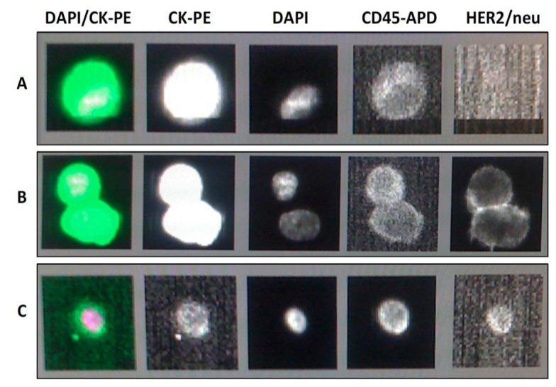
Figure 1.
Typical appearance of isolated cells using the CellSearch® method. DAPI: fluorescently stains nuclear material; CK-PE: detects presence of cytokeratin 8, 18, 19; CD45-APD: detects presence of CD45. (a) HER2 negative CTC: this cell is a CTC evidenced by CK positivity (2nd image from left) and DAPI positivity (3rd image) and CD45 negativity (4th image). HER2 immun(ofluorescence is negative (5th image); (b) HER2 positive CTC: CK is again positive, DAPI positive and CD45 negative consistent with this being a CTC. HER2 immunofluorescence is positive (5th image from left); (c) Lymphocyte: This cell is not a CTC as it is negative for CK (2nd image from left, compare with the two images above). DAPI positivity and CD45 positivity indicates this is a lymphocyte.

