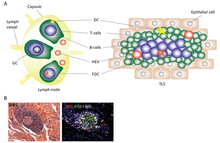
Figure 1.
Histological similarities and structural differences between lymph nodes and TLS. (A) Both lymph nodes and TLS contain the same cell populations and high endothelial venules (HEV). On the left, a schematic of lymph node structure highlighting B and T cell zones is shown. Each zone contains resident cell populations that upon antigen presentation by follicular dendritic cells (FDC) or DC, and subsequent activation, undergo clonal expansion. Expanded B cell populations form a germinal center (GC). On the right, a TLS schematic showing individual cells aggregating which mimics lymph node histological structure is shown. B cells in this case will also clonally expand and form germinal centers after antigenic stimulation. Structural differences are highlighted; lymph nodes are encapsulated and connected to the lymphatic system via afferent and efferent lymph vessels while a TLS forms within a chronically-inflamed tissue and lymph vessel formation may eventually occur [6]; (B) Tissue specimen of TLS structures seen in tuberculosis infection. The left is an H&E stain; the right is an immunoflourescence image staining for CD3+ T cells and CD21+IgD+ B cells [7].

