Figure 1.

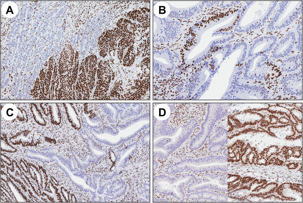
Examples of the different MMR protein staining patterns. A) clonal loss, B) intraglandular loss, C) co-existence of clonal and intraglandular loss and D) compartmental loss with different patterns in two separate tumor blocks.

Examples of the different MMR protein staining patterns. A) clonal loss, B) intraglandular loss, C) co-existence of clonal and intraglandular loss and D) compartmental loss with different patterns in two separate tumor blocks.