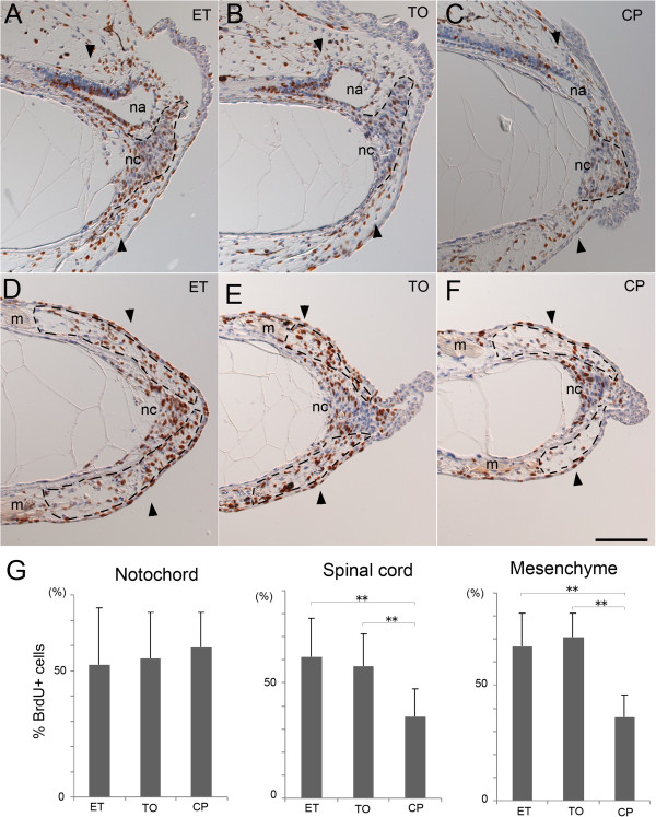
Figure 4.

Cyclopamine affects cell proliferation in the regenerating tail. Tail-amputated tadpoles maintained in the presence of the indicated compound were incubated with BrdU for 12 h before fixation. (A-F) Immunohistochemical detection of proliferation cells at day 2.5. BrdU-incorporated cells (brown) were detected on sagittal (A-C) or frontal (D-F) section. Un-labeled nuclei were counter-stained with hematoxylin (blue). Dashed lines indicate the shapes of the regenerating notochords (A-C) and the mesenchymal regions (D-F) containing the myoblasts. nc, notochord. na, neural ampulla of the regenerating spinal cord. m, muscle. A pair of arrowheads marks the amputation plane. Bar, 100 μm. (G) Proliferation rate of cells in the regenerating tail. Mean rate of the BrdU-labeled cells was determined for the indicated tissue and shown with standard deviation. Detail is shown in Table 3. **, p-value <0.01.
