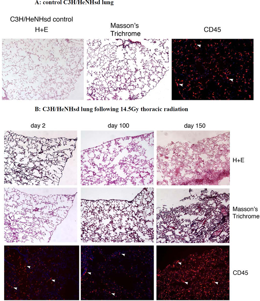
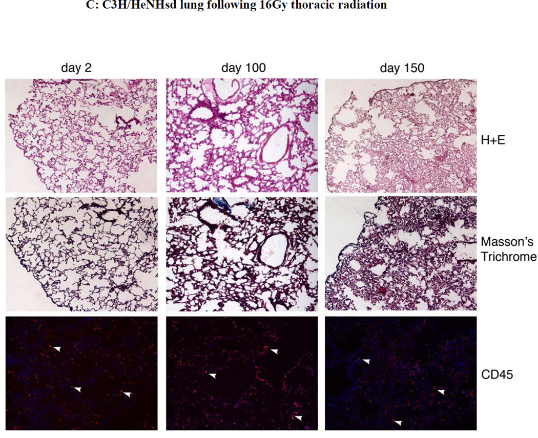
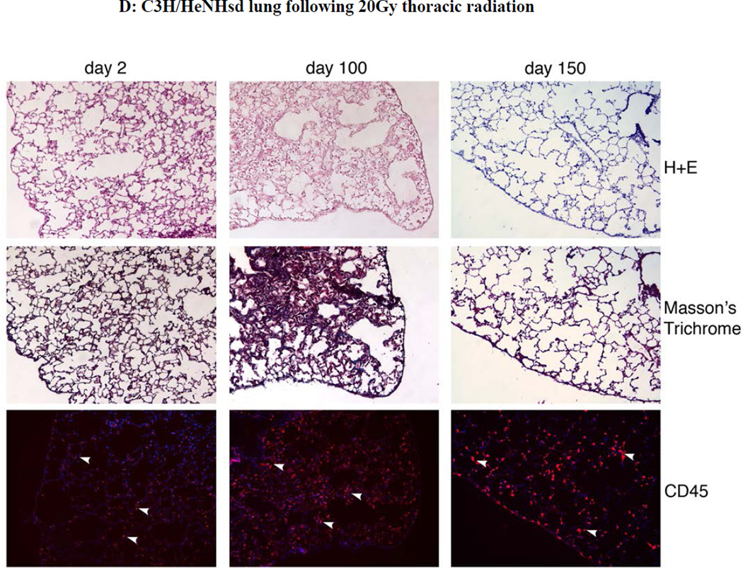
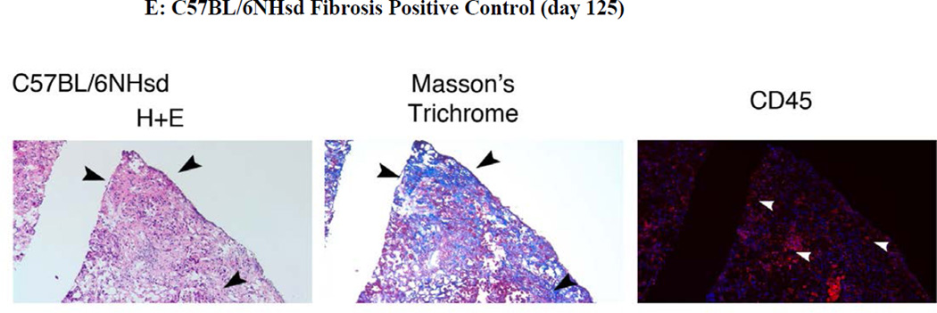
Figure 5. Absence of pulmonary fibrosis in lungs of thoracic irradiated C3H/HeNHsd mice.
Frozen sections removed at day 2, 100, or 150 from lungs of: A) unirradiated C3H/HeNHsd control mice; B) C3H/HeNHsd mice thoracic - irradiated to 14.5; C)16 Gy; or D) 20Gy; or E) 20Gy thoracic-irradiated C57BL/6NHsd mice were stained with hematoxylin and eosin, Masson’s trichrome (for collagen) or immunostained for CD45 at serial times after irradiation. Sections were collected from C3H/HeNHsd mouse lungs at day 2 (acute phase), day 100 (latent period) and day 150 (late phase). No detectable fibrosis or increased collagen was observed in control unirradiated, acute, latent or late phase lung tissue from C3H/HeNHsd mice after any irradiation dose. C57BL/6NHsd mice showed late phase fibrosis at day 150 (E). Increased collagen (black arrows) in Masson’s trichrome stained sections (E) and increased numbers of red stained CD45+ inflammatory cells (white arrows) in peri-vascular distribution was seen in C57BL/6NHsd lungs in the late phase post-irradiation (E). Masson’s trichrome staining of lungs from irradiated C3H/HeNHsd mice at all times was similar to control unirradiated lungs, indicating no detectable fibrosis. Low level immunostaining for CD45 cells (white arrows) in C3H/HeNHsd lungs was similar in control unirradiated, and irradiated lungs at all phases. All images 10 x.


