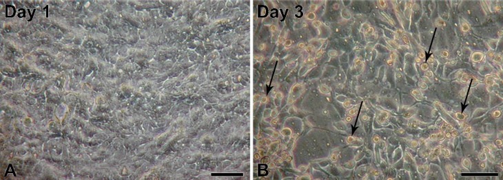
Figure 1.
Micrographs of normal porcine urothelial (NPU) cell cultures of the XIII passage on porous membranes on a first (A) and third (B) day after seeding at a density of 2 × 10⁵ viable cells/cm2. Note the fast cell attachment on the first day, where NPU cells are almost confluent, and an increased cell desquamation on a third day after seeding, where numerous rounded and detached NPU cells can be seen (arrows). Scale bars: 100 µm.

