Figure 5.
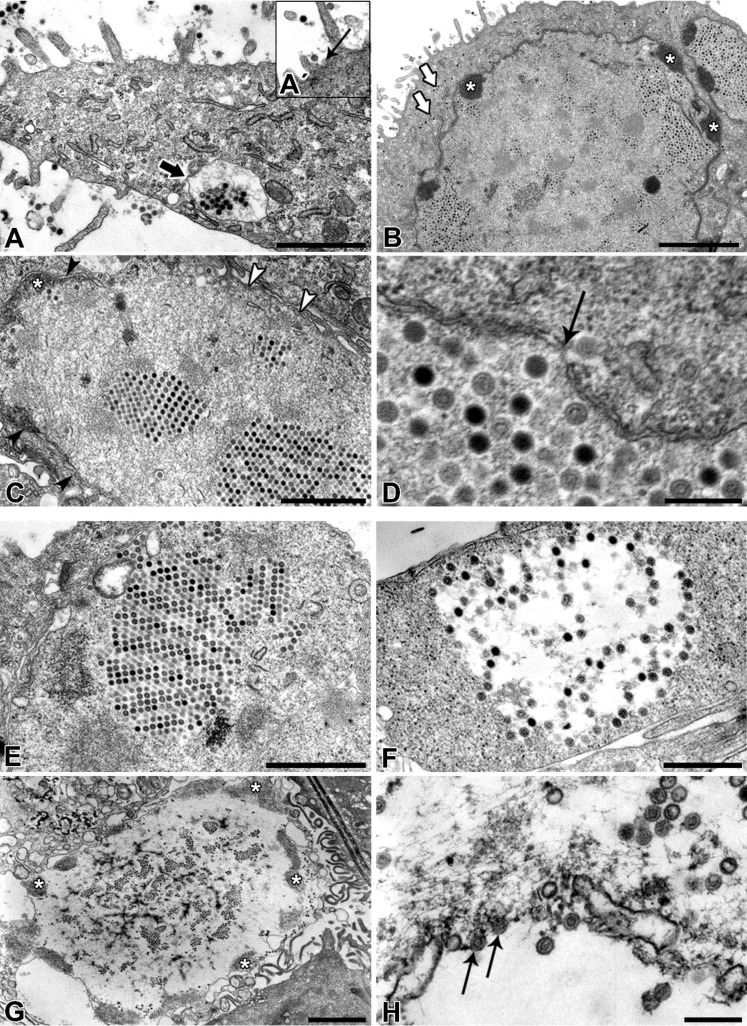
Transmission electron micrographs of PAdV-SVN1 in NPU cells isolated from porcine urinary bladder. PAdVs enter the NPU cells by endocytotic uptake (arrow on A´). After the endocytosis, the virions are found in late endosomes (A) or sporadically scattered in the cytosol (arrows on B). After the cell internalization, the virions are found in the nucleus, organized in larger paracrystalline arrays or spread throughout the nucleus in smaller clusters (B). Commonly the condensed chromatin at the nuclear envelope of infected NPU cells is noted (asterisks on B and C). The possible mechanism of nuclear escape includes the separation of nuclear envelope and merger of the nucleus full of virions with the cytosol (black arrowheads on C indicate the nuclear envelope, white ones the plasma membrane), or maybe even the individual release of virions through the nuclear pore (arrow on D). After re-entry to the cytosol, the virions are found both, in the large paracrystalline arrays (E) or randomly distributed throughout the cytosol (F), with frequently observed degradation of the cytoplasm at the site of their presence (F). At this point, the initiation of NPU cell necrosis can be observed. With the progress of cell lysis, the cell detaches from the epithelium, i.e., urothelium (G); the nucleus disintegrates and condensed chromatin is moved near the plasma membrane (asterisks on G). At the final stage of necrosis, virions leave the cell through the lysed plasma membrane (arrows on H). Scale bars: (A, C and E) 1 µm; (B) 20 µm; (D and H) 200 nm; (F) 500 nm, (G) 2 µm.

