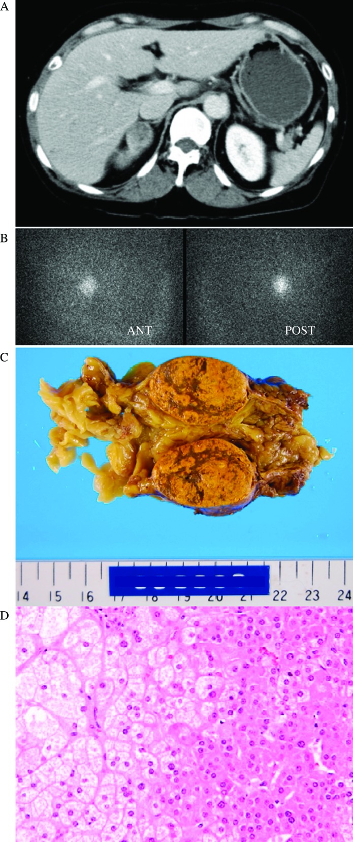
Figure 2.
Abdominal computed tomography showing a 31×24 mm low-density mass within the right adrenal gland with mild enhancement post-contrast (A) that was also positive for [131I]adosterol imaging (B). Cut sections of the yellow adrenocortical adenoma (30×30×20 mm, 25 g nodule) with the atrophic right adrenal gland (C). Pathological evaluation of the right adrenal mass indicated diagnostic characteristics of a typical adrenocortical adenoma with Cushing's syndrome (D).

 This work is licensed under a