Fig. 2.
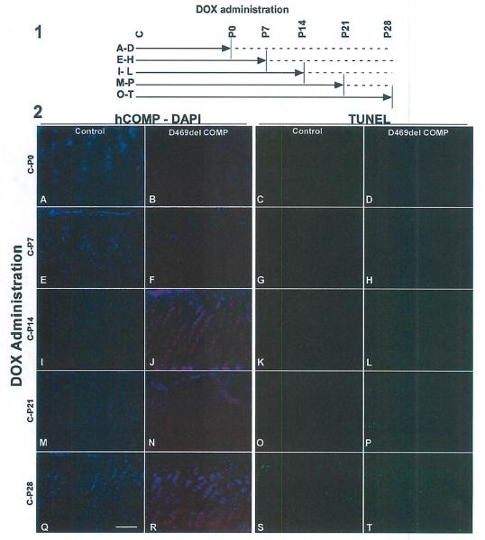
Chondrocyte cell death increases with duration of D469del-COMP expression. Panel 1: (A–T) DOX was administered for weekly intervals according to the schematic. Solid line indicates DOX administration to mice (500 ng/ml); dotted line indicates no DOX. All treatments were started at conception (shown as “C”)and continued until P0, P7, P14, P21, or P28. Growth plates from all animals were analyzed at P28. Panel 2: D469del-COMP (red) and DAPI (blue-nuclei) staining of D469del-COMP (B, F, J, N, and R) and control C57BL/6 (A, E, I, M, and Q) growth plates are shown. Very little intracellular D469del-COMP was observed with DOX administration from birth to P7. TUNEL staining of D469del-COMP (D, H, L, P, and T) and control C57BL/6 (C, G, K, O, and S) growth plates are shown. Cell death increased dramatically in D469del-COMP growth plates after 2 weeks of DOX (L, P, and T) compared to controls (K, O, and S). Bar = 500 μm.

