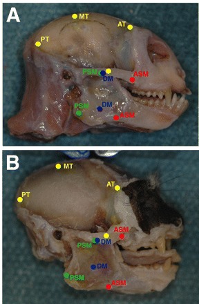
Fig. 4.

Sagittal view of Callithrix jacchus (A) and Saguinus oedipus (B) skulls with the masseter and temporalis muscles removed. The origin and insertion points of the anterior (AT), middle (MT) and posterior temporalis (PT; yellow circles) as well as the anterior superficial (ASM; red circles), deep (DM; blue circles) and posterior superficial (PSM: green circles) masseter are shown. Markings were made on the skulls as muscles were removed to approximate muscle paths.
