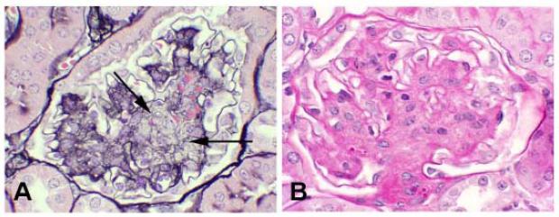
Figure 2.
Morphologically advanced murine diabetic nephropathy demonstrating prominant mesangiolysis. A. There are broad areas of lucency (arrows) within expanded mesangial regions. Mesangiolysis is most easily recognized with a Jones’ silver methenamine stain, in which the normally homogeneous and compact silver staining (black) matrix (shown in Figure 1A) is disrupted as indicated by areas of lucency and/or spongiform appearance. B. In contrast, a PAS stain shows equivalent mesangial expansion, but the demarcation of cellular and matrix components and lytic regions is less distinct with this stain as compared to the Jones’ stain.

