
Figure 3. Reversibility of Dex effects in A549 cells.
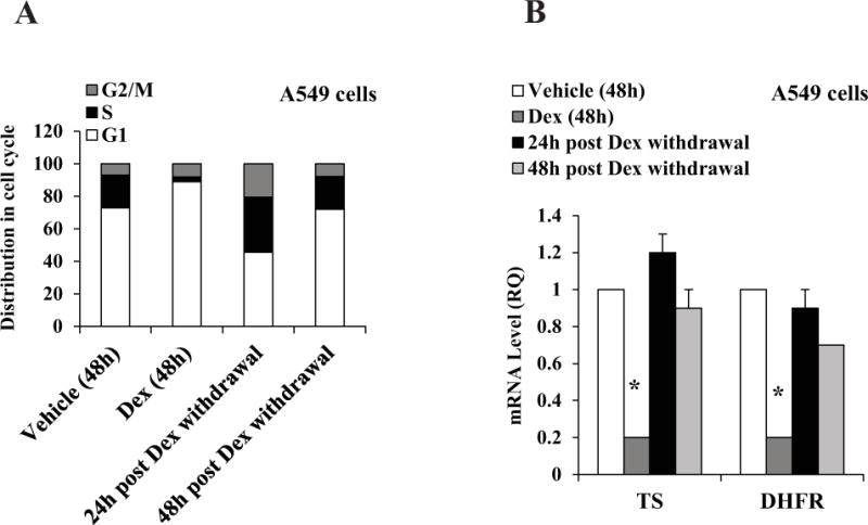
A549 cells were plated at 20 percent confluence in media containing charcoal-stripped serum for 24 h for hormone depletion. Cells were then treated with either vehicle (ethanol) or Dex (100 nM). After 48 h, Dex was removed by washing the cells twice and replacing with fresh media. Cells were cultured for additional periods of 24 h and 48 h. Cells were harvested for flow cytometry analysis (Panel A). The experiments were repeated at least three times. Within each experiment, the percent error for replicate samples was < 10 percent. The P values for Dex induced changes noted in the text were < 0.0001. In parallel, cells were harvested for mRNA measurement by real time RT-PCR (Panel B). * P < 0.01. All of the mRNA measurements were carried out using biological triplicate samples.