
Figure 6. Differential effects of Dex on growth inhibition by pemetrexed in A549 vs. H1299 cells.
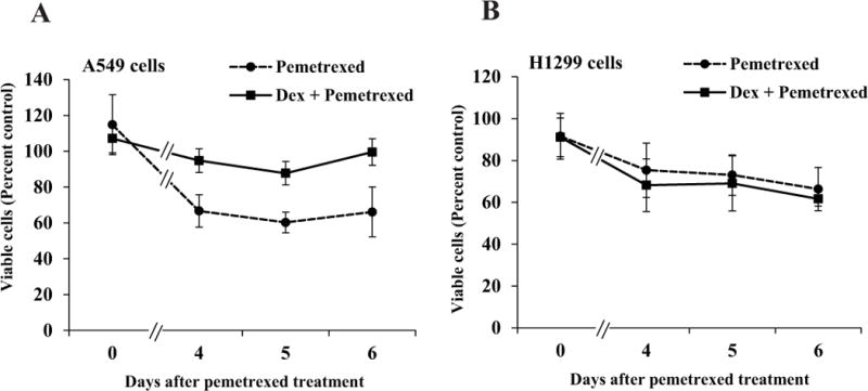
A549 cells (Panel A) and H1299 cells (Panel B) were plated in six replicate wells in 96 well plates in media containing charcoal-stripped serum as described under Materials and Methods for MTT assays. 24 h after plating, the cells were treated with vehicle (ethanol) or Dex (100 nM) for 72 h – 96 h. The cells were exposed to pemetrexed (5 μM) or vehicle (water) for a 24 hour window in the midst of the Dex (or vehicle) treatment. MTT assays were performed on the indicated days, beginning with the day of pemetrexed treatment (Day 0). At each time point, the ratio of the absorbance for pemetrexed treatment to its vehicle control was used to determine the percentage of viable cells.