
Figure 1.
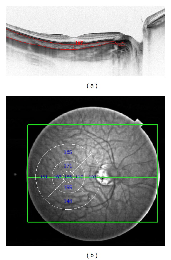
(a) Swept source OCT in a healthy volunteer. The red horizontal lines were drawn automatically at the inner and outer choroidal boundaries. The vertical line was drawn manually, to measure central choroidal thickness. (b) Swept source OCT fundus view. The circle and numbers are drawn automatically and represent automated thickness and volume measurements, which were performed in 9 segments according to early treatment diabetic retinopathy study.