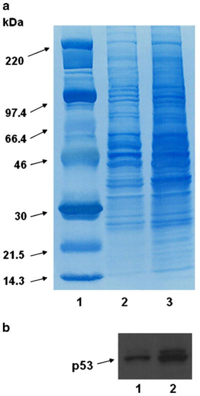
Fig. 1.

Differences in antigen release from tumor cells by freeze thaw (F/T) alone and freeze-thaw + 15 sonication (F/T + S). In order to determine differences in protein content, lysate samples from identical numbers of starting cells were electrophoresed on a SDS-PAGE mini-gel system and transferred onto nitrocellulose membranes. After an initial screen, two techniques (F/Tand F/T + S) resulting in relatively greater protein liberation from cellular lysates were compared against one another. Greater intensity of staining in the lanes loaded with lysate subject to F/T + S implied greater antigen release by this method, an observation confirmed by increased release of the defined tumor Ag (p53). (a) Coomassie Blue staining: Lane 1: Molecular weight ladder; lane 2: F/Talone; lane 3: F/T + S. (b) Western blot utilizing an anti-p53 antibody shows increased release of this protein: Lane 1: F/Talone; lane 2: F/T + S.
