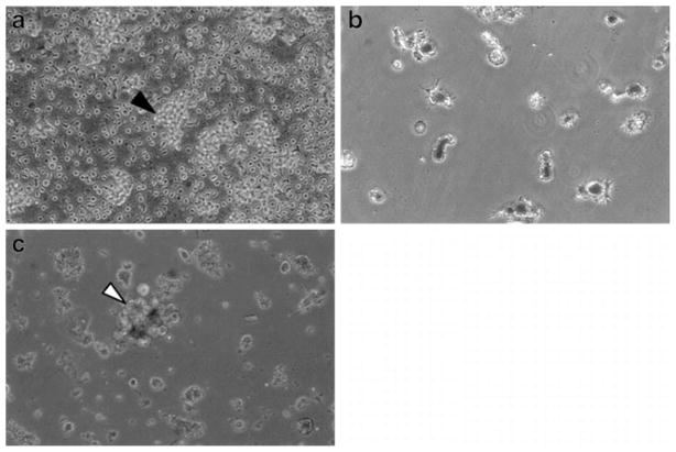
Fig. 5.
Phase contrast microscopy of components of DC/TIL co-culture: (a) TIL were isolated and expanded using CD3/CD28 Dynabeads®; black arrow indicating conglomeration of expanded TIL. (b) PBMC-derived DC were loaded with TA containing-80KDa PLGA NP; cells demonstrating characteristic dendrites and visible NP associated with the cell surface and surface invaginations. (c) Co-culture of freshly obtained autologous antigen loaded-DC with TIL; white arrow depicting representative DC/TIL complex.

