Figure 3.
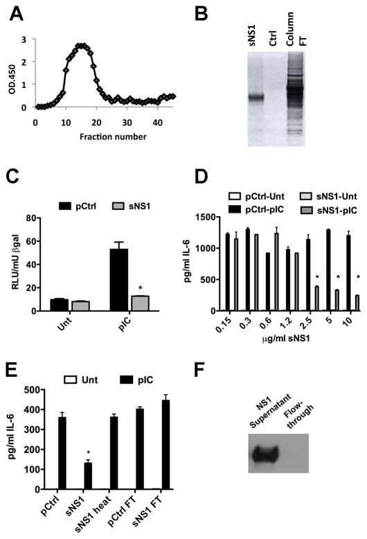
Incubation with purified sNS1 inhibits TLR3 signaling in HeLa cells. (A) NS1-specific ELISA on immunoaffinity purification fractions. (B) Silver stain on SDS PAGE gel of purified sNS1, the purification control (pCtrl) and the flow through fraction during the purification. (C) HeLa cells were transfected with IFNβ reporter construct and β-gal expression construct and incubated with 4μg/mL purified sNS1 or an equal volume of pCtrl for 16h prior to stimulation with pIC. Control for these experiments was supernatant from 293T vector control cells that underwent a purification process identical to that of the sNS1-containing supernatants (pCtrl). Asterisks denote statistically significant differences between pCtrl and sNS1 pretreated pIC-stimulated cells.
(D) Dose response curve. Varying concentrations of purified sNS1 or equal volume of pCtrl was added to HeLa cells in full serum media and incubated for 16h before 8h of pIC treatment and subsequent IL-6 ELISA analysis of supernatants. Asterisks denote statistically significant differences between pCtrl and sNS1 pretreated pIC-stimulated cells.
(E) IL-6 secretion was measured from pIC treated HeLa cells following incubation with pCtrl, 5μg/mL sNS1, 5μg/mL sNS1-heat or equal volumes of the flow through fraction generated during the purification process for either control cell supernatant or NS1-expressing cell supernatant. (F) Western blot confirming depletion of sNS1 in the flow through fraction used in Fig. 3E. Asterisks denote statistically significant differences between pCtrl and NS1 pretreated cells, stimulated with pIC.

