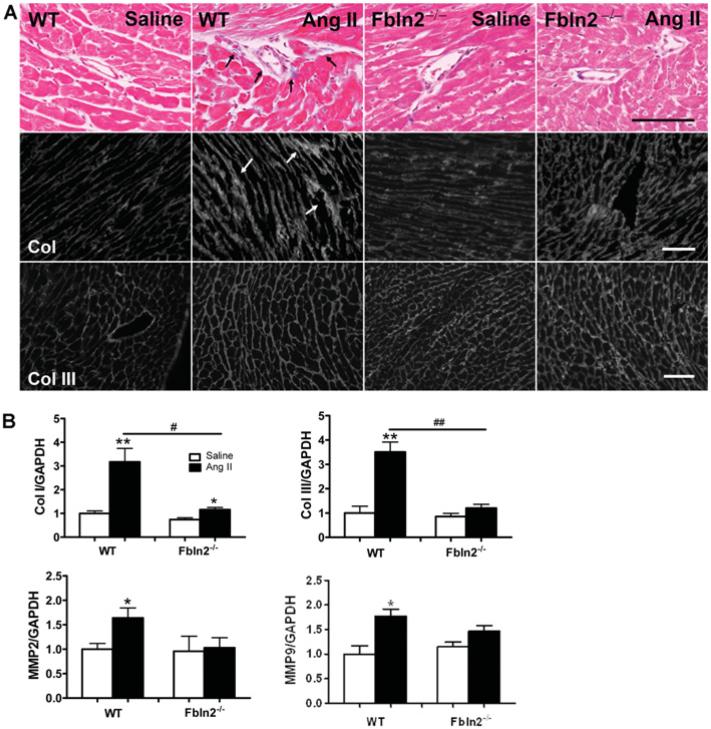
Figure 3. Assessment of ECM remodelling and tissue fibrosis in LV myocardium.
(A) Masson's Trichrome (Ma-Tri) staining and immunohistological staining (Col I and Col III). In Masson's Trichrome, blue colour indicates the area of collagen fibres (see arrows), which is exclusively seen in the perivascular area. There was no noticeable increase in tissue collagens by AngII in either WT or Fbln2−/− . However, by immunostaining, interstitial deposition of Col I was markedly increased in AngII-treated WT (see arrows) compared with AngII-treated Fbln2−/− . There was no significant difference in Col III protein deposition in all four groups. Also note that the size of each myocyte was markedly enlarged in AngII-treated WT compared with other three groups, consistent with Figure 2(B). Scale bar, 100 μm. (B) Col I, Col III, MMP-2 and MMP-9 mRNA levels by qRT–PCR. The mRNA levels of saline-treated WT were arbitrarily set at 1. WT (saline, n = 10; AngII, n = 15) and Fbln2−/− (saline, n = 10; AngII: n = 15). #P < 0.05 and ##P < 0.01. *P < 0.05 and **P < 0.01 compared with corresponding saline-treated group.

