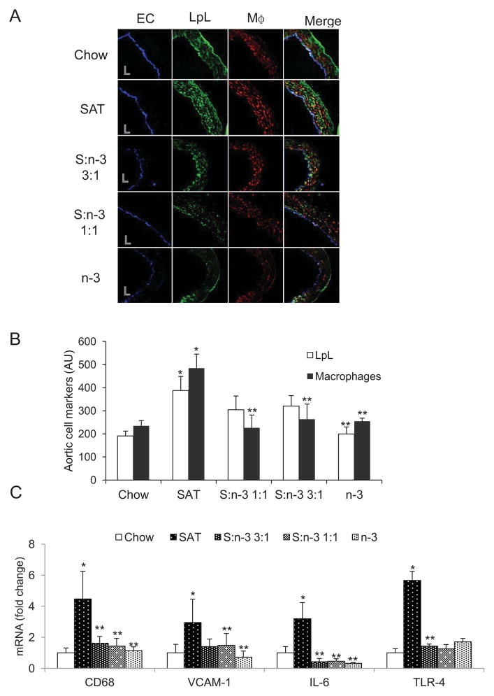
Figure 2.
Effects of diet on LpL, macrophages and inflammatory markers in proximal aorta in LDLR−/− mice. (A). Representative images of proximal artery sections stained for endothelial cells (EC), LpL and macrophages (MΦ) in LDLR−/− mice that were fed the specific diets for 12 weeks. “L” indicates lumen. (B). Immunofluorescence of aortic LpL (open bars) and macrophages (filled bars) quantitated for each group. Mean±SE, n=3–4. (C) mRNA analyses of arterial pro-inflammatory markers. Proximal aorta homogenates of LDLR−/− mice (n=5) were analyzed for mRNA of CD68, VCAM-1, IL-6 and TLR-4 in each group. Data are expressed as mean±SE. *, SAT vs. chow; **, vs. SAT, p<0.05.

