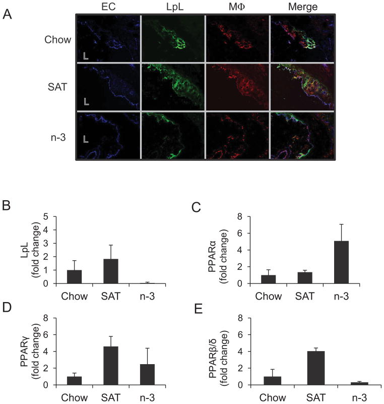
Figure 3.
Effects of diet on LpL and macrophage localization and LpL/PPAR mRNA at the aortic origin in LDLR−/− mice. (A) Images shown are the colocalization of aortic EC, LpL, and macrophages (MΦ) in LDLR−/− mice fed a chow, SAT, or n-3 diet for 12 weeks. L=lumen. Aortic macrophages were collected by LCM and pooled (n=3–5)for measuring mRNA expression of LpL (B), PPARα (C), γ (D), and β/δ (E) with triplicate runs (mean±SE) in each group as described in Methods.

