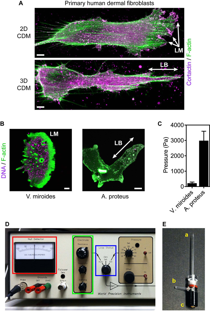
Figure 1.
Direct intracellular pressure measurements in migrating cells. (A) Maximally projected confocal stacks of primary human dermal fibroblasts migrating on (upper panel) 2D and in 3D (lower panel) cell-derived matrix (CDM) stained for filamentous actin (F-actin) with rhodamine-phalloidin (green) and the lamellipodia marker cortactin (magenta). Fibroblasts can migrate on 2D surfaces using low-pressure lamellipodia (LM) and in 3D matrix using high-pressure lobopodia (LB). Scale bars, 5 µm. (B) F-actin structures in the protists Vanella miroides (lamellipodia) and Amoeba proteus (lobopodia). Cells were stained with SYBR green (DNA, magenta) and rhodamine-phalloidin (F-actin, green). Scale bars 10 µm in (A) and 50 µm in (B). (C) The intracellular pressure of lamellipodial V. miroides and lobopodial A. proteus. (D) The control panel of the 900A micropressure system (WPI). (E) Micropipette and microelectrode holder labeled for the microelectrode tip (a), air connection (b) and the microelectrode connection (c).

