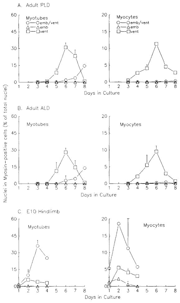
Fig. 3.
Frequency of MHC-positive myocytes and myotubes in double-labeled adult and fetal primary myogenic cultures from various muscles. (A) Adult posterior latissimus dorsi (PLD)-, (B) Adult anterior latissimus dorsi (ALD)-, and (C) E10 hindlimb-derived cultures. Duplicate cultures were labeled and data obtained as in Fig. 2. Error bars show standard deviation; where there are no error bars, the symbols are bigger than the standard deviation. Values decrease due to continued proliferation of nonmyogenic cells. emb/vent, coexpression of embryonic and ventricular MHCs; emb, expression of embryonic MHC; vent, expression of ventricular MHC.

