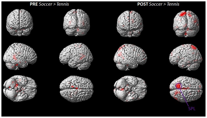
FIGURE 5.
Group activation maps showing activated brain regions in condition “PRE Soccer > Tennis (left side) and POST Soccer > Tennis (right side).” All regional activations above initial significance threshold P < 0.05 (FEW corrected) and extent (kE) of 30 voxels are depicted on a rendered MNI brain. SPL = superior parietal lobule.

