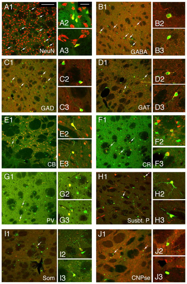
Figure 2. Most MGE Cells Adopted an Inter-neuronal Fate when Transplanted into the Adult Striatum.
(A) Four weeks after transplantation, most MGE cells expressed the mature neuronal markers NeuN (75% ± 6%).
(B–D) Most MGE cells also expressed markers related to GABA storage or synthesis such as GABA (B, 75% ± 4%), GAD (C, 60% ± 11%), and GAT (D, 50% ± 9%).
(E–I) Some transplanted MGE cells also expressed interneuron subtype markers CB (E, 24% ± 7%), CR (F, 8.3% ± 0.5%), PV (G, 0.5% ± 0.5%), Substance P (H, 6% ± 2%), and Somatostatin (I, 1% ± 0.5%).
(J) One quarter of the MGE cells transplanted into the striatum (25% ± 4%) expressed the oligodendrocyte marker CNPase. Arrows point to examples of MGE cells that stained positive for each marker, and insets for each panel (labeled 2 and 3) show higher power examples of immunopositive cells.
Scale bar represents 30 μm in (A1) and applies to lower-power images shown in (A1)–(J1); scale bar represents 10 μm in (A2) and applies to all higher-power inset images (A2–J2 and A3–J3).

