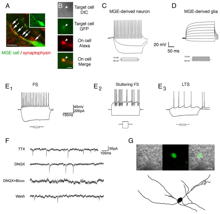
Figure 3. MGE Cells Transplanted into the Striatum Displayed the Basic Membrane Properties Characteristic of Mature Forebrain Interneurons and Showed Evidence of Synaptic Integration.
(A) 67% of the MGE cells (green) expressed the synapse marker synaptophysin (red) along their processes (white arrows).
(B) We obtained whole-cell patch-clamp recordings from transplanted cells up to 20 weeks after transplantation. Alexa-594 (red) was included in the fill solution of the glass microelectrode to confirm that recordings were obtained from targeted GFP-expressing MGE transplant cells.
(C) Most MGE cells (37/44) fired repetitive nonaccommodating action potentials with large after hyperpolarizations as is common for MGE-derived interneurons. (D) A small percent of the transplanted MGE cells that were recorded from (n = 7/44) did not exhibit neuronal membrane properties and did not fire action potentials when stimulated with a series of depolarizing currents, consistent with glial cell identity.
(E1) Most MGE neurons were fast spiking (FS) cells and did not fire rebound action potentials after hyperpolarization.
(E2) The FS population included stuttering FS cell subtypes.
(E3) The second most common cell type was low-threshold-spiking (LTS) cells. These cells displayed much lower spiking frequencies than did FS cells and displayed two or more rebound spikes in response to hyperpolarization.
(F) Transplanted cells demonstrated both AMPA and GABA-mediated spontaneous synaptic events.
(G) A transplanted MGE cell filled with Texas red-dextran demonstrated the branched aspiny processes that are characteristic of striatal interneurons.
Scale bars represent 3 μm in (A), 5 μm in (B), and 10 μm in (G).

