
Figure 7. MGA Cells Transplanted in the Subthalamic Nucleus.
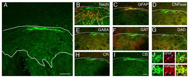
(A) MGE cells transplanted into the subthalamic nucleus (STN) survived but did not migrate from the site of injection and did not mature into neurons.
(B–K) None of the MGE cells that were transplanted into the STN expressed the neuronal marker NeuN (B), the inhibitory interneuron marker GABA (E) or GAD (G), or the calcium sequestering proteins CR (H) or CB (I). Most MGE cells transplanted into the STN expressed the astrocyte marker GFAP (75% ± 5%, C and J) or the oligodendrocyte marker CNPase (30% ± 9%, D and K). A small percentage of MGE cells in the STN expressed the GABA transporter GAT1 (13% ± 5%, F).
Scale bars represent 25 μm in (A); 25 μm for (I) and (B)–(H); and 5 μm for (K) and (J).