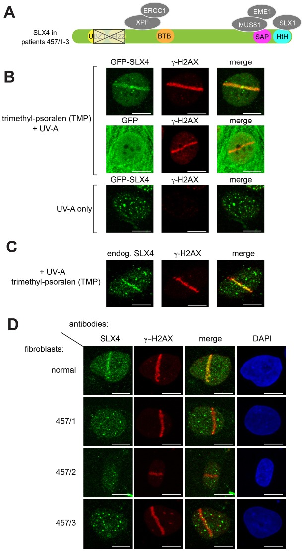
Fig. 1.
Recruitment of SLX4 to sites of localized DNA damage induced by PUVA. (A) Diagram showing SLX4 domain organization and associated nucleases. Siblings 457/1, 457/2 and 457/3 are FA patients with a deletion (outlined in black) removing all of the second of the tandem SLX4 UBZ domains and part of the first. BTB; Broad-complex, Tramtrack, Bric-a-brac domain: SAP; SAF-A/B, Acinus and PIAS motif: HtH; helix-turn-helix motif. (B) U2OS cells stably expressing GFP–SLX4 (upper and lower panels) or GFP only (middle panels) were incubated (or not) with trimethyl-psoralen (TMP; 20 µM, 60 min) and subjected to subnuclear micro-irradiation using a 355-nm UV-A laser. Cells were fixed and subjected to indirect immunofluorescence analysis with antibodies against GFP or γ-H2AX. (C) As for B except the localization of endogenous SLX4 (endog.) in U2OS cells was examined. (D) Cells from FA patients 457/1, 457/2 and 457/3 or normal human fibroblasts were treated as in B and endogenous SLX4 localization was analyzed by using indirect immunofluorescence. Scale bars: 10 µm.

