Fig. 2.
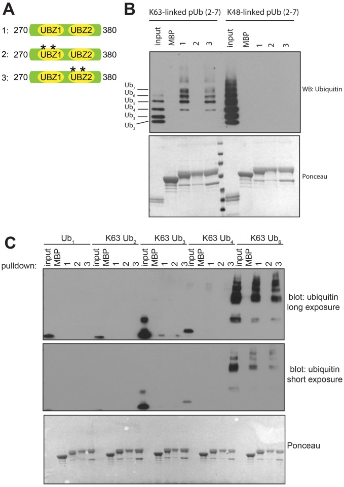
Testing the ability of the SLX4 UBZ domains to bind to ubiquitin. (A) Diagram of the MBP-tagged SLX4 fragments used in ubiquitin-binding assays. Asterisks denote cysteine to alanine mutations at Cys 296 and Cys 299 in UBZ-1 (fragment 2) or Cys 336 and Cys 339 in UBZ-2 (fragment 3). (B) The fragments shown in A or MBP alone were immobilized on amylose–agarose and incubated with K48-linked or K63-linked poly-ubiquitin (pUb, 2–7) chains. Pulldowns were subjected to SDS-PAGE and immunoblotted (WB) with anti-ubiquitin antibodies. The bottom panel shows a Ponceau staining of the membrane performed prior to blotting. (C) Same as for B, except that mono-ubiquitin or K63-linked poly-ubiquitin chains of the indicated length were used with fragments 1–3.

