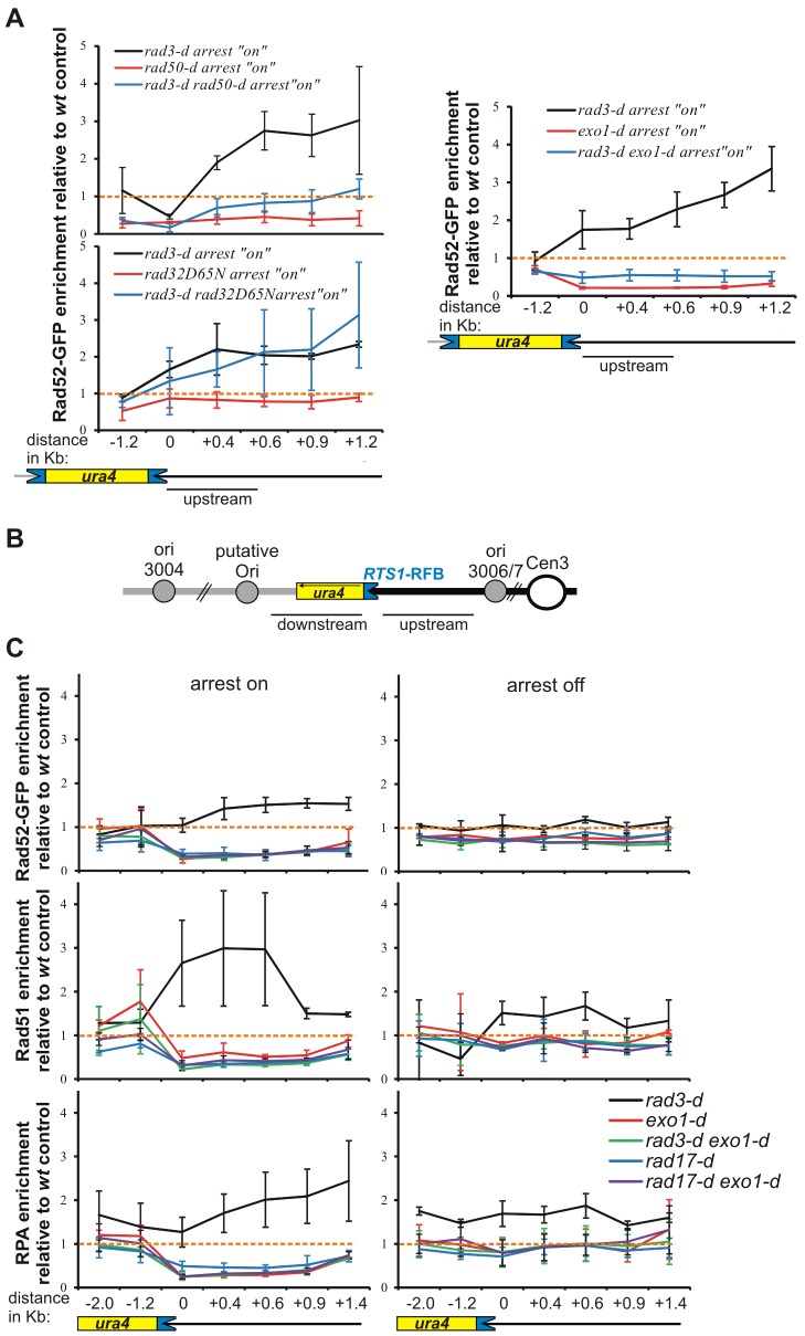
Fig. 2.
Rad3ATR and Rad17 regulate Exo1-dependent recruitment of ssDNA-binding proteins at collapsed forks. (A) Rad52–GFP enrichment relative to wild-type control (wt) at the RuraR locus in the indicated strains, as described for Fig. 1D. Data show the mean±95% CI (three independent experiments). Schematics are as described for Fig. 1A. (B) Schematic representation of the uraR locus, as described for Fig. 1A. (C) Relative enrichment of Rad52–GFP (upper panels), Rad51 (middle panels) and RPA (lower panels) relative to the wild-type control (wt) for indicated strains, as described for Fig. 1D. ChIP followed by qPCR was performed on the indicated uraR strains after 40 hours of growth either with or without thiamine (arrest ‘off’ and ‘on’, respectively). Orange dotted lines, relative enrichment of 1. Data show the mean±95% CI (three independent experiments).

