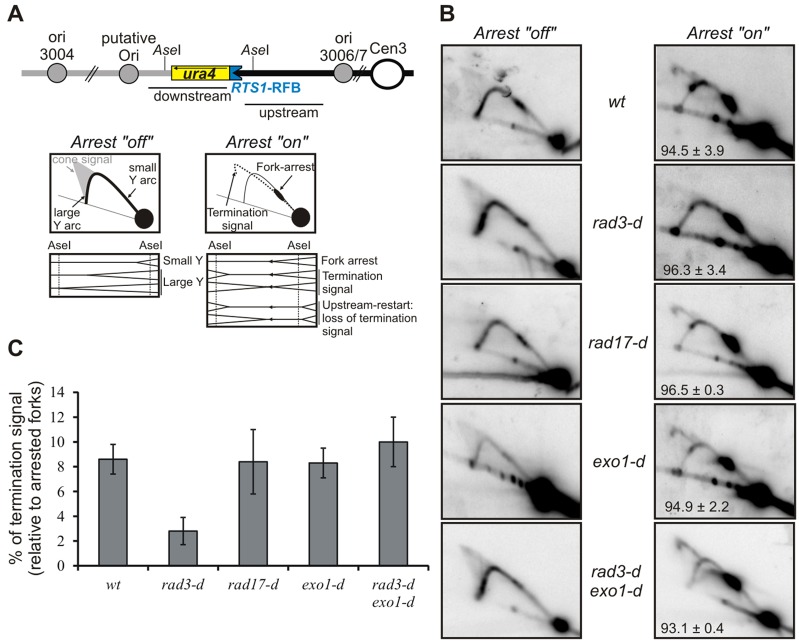
Fig. 3.
Exo1-dependent fork resection is regulated by Rad3ATR and Rad17. (A) Schematic representation of the uraR locus, as presented in Fig. 1A. Panels show diagrams of replication intermediates within the Ase1 restriction fragment as analysed by 2DGE under the indicated conditions. (B) Analysis of replication intermediates by 2DGE from the indicated strains after growth for 24 hours in medium with or without thiamine (fork arrest ‘off’ and ‘on’, respectively). wt, wild-type control. Numbers indicate the percentage of forks arrested by the RTS1-RFB (±s.d.) (C) Quantification of the termination signal from B in the indicated strains. Data show the mean±s.d. (three independent experiments).

